embodiment etudes

short somatic studies applied to yoga

Month: March, 2019

the reproductive organs

by julee snyder

2nd chakra

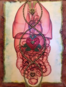
The territory of the second chakra, the reproductive organs can add lots of layers to grounding us to the earth and countering the reach of the upper extremities into space.  Let’s look at the different structures to begin our explorations.

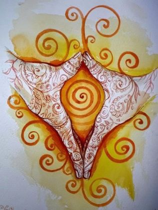
Female Reproductive System

The female reproductive system consists of the ovaries, fallopian tubes, uterus, vagina, vulva and mammary glands.  This system produces the female sex cell, the ova, and creates an environment for fertilization and subsequent development of the fetus.

uterus

The uterus is in the center of the lower portion of the pelvic bowl slightly above the pubic arch and at the same level as the coccyx.  It lies atop of the bladder.  It is a small, muscular, pear-shaped organ.  In its non-pregnant state it measures approximately three inches in length, two inches in width and one inch thick.  It has very strong, but stretchable muscular fibers.  Most of its outer surface is covered by peritoneum; it is sub peritoneal.  The uterus is moveable, but held into place by peritoneal ligaments.

The function of the uterus is to hold the embryo during pregnancy and expel the fetus at birth through powerful contractions.  Each month the inner lining of the uterus undergoes changes in preparation for a fertilized egg.  If fertilization does not occur, the lining is sloughed off in a hemorrhagic process known as menstruation.

ovaries

The ovaries lie above and on either side of the uterus in the folds of the broad ligament with e mesentery attachment called the mesovarium.  The ovarian ligament anchors each ovary to the uterus.  They are partially encircled by the fallopian tubes, which are also nestled in the broad ligament.

The ovaries contain between 40,000 and 400,000 immature ova.  Alternating between the two ovaries, one ova develops and is discharges every 28 days during childbearing years.  The ovaries secrete estrogen and progesterone.


Male Reproductive System

The testicles and penis lie externally to the lower pelvic cavity.  Internally the bladder is in front of the prostate gland followed by the seminal vesicles and rectum.  The vas deferen tubes connecting the testicles to the seminal vesicles follow above and around the bladder.

The testicles, penis, prostate gland and seminal vesicles form the male genital system.  The urethra located in the penis connects the genital and the urinary systems.   The genital system produces and stores sperm and secretes semen for reproduction.  The genitourinary system ejaculates seminal fluid and eliminates urine from the body.

testes

The testes are contained in a pouch of skin called the scrotum.  They are located externally below the pubic symphasis and in front of the perineum.  The testes, like the ovaries, develop along the back wall of the abdominal cavity and descend into the scrotum just before birth.

There are two testicles contained in the scrotum.  The testicle, a slightly flattened, oval-shaped gland, is about the size of a walnut.  The tunica albuginea – the outer cavity of the testicle – has a tough, fibrous, white surface, which is comparable to the outer coat of the eyeball.  Internally, the testes are divided into about 250 elongates pyramidal compartments, each containing a lobule formed from two or more seminiferous tubules.  These seminiferous tubules form sperm.  The epididymis, a coiled C-shaped structure is located along the upper posterior border of the testicle and is a storehouse for sperm cells.

The testes produce, store and distribute spermatazoan and act as an endocrine gland secreting testosterone.

seminal vesicles

The seminal vesicles are locates in front of the rectum along the lower, posterior surface of the bladder and behind the prostate gland.  The seminal vesicles are two small, convolutes pouches which secrete a viscous liquid that combines with sperm from the testicles to produce semen.  This liquid also contains nutrients that support sperm metabolism.

prostate gland

The prostate gland is located behind the pubic symphasis just below the bladder.  The prostate gland encircles the tube of the urethra with two lateral lobes and a median lobe.  Smooth muscle is interwoven throughout the gland to facilitate emission of semen through the ejaculatory ducts that converge within the prostate gland.  The prostate gland secretes an alkaline substance that combines with the seminal fluid to protect the sperm from acid present in the male urethra.

vas deferens

The vas deferens tubules transport sperm from the testes to the urethra.  The vas deferens leave the epididymis ascending through the inguinal canal into the lower pelvis where they loop up and around the bladder before descending into the seminal vesicles behind the prostate.  There it merges with the seminal vesicle duct and forms the ejaculatory duct.


Embodying the Reproductive System

When embodying the reproductive system, energize each organ with breath.  Move each organ on its axes in all planes.  Relate each organ to the extremities: feet, brain, hands.  Find the triangle of uterus/prostate to gonads.  Relate the uterus/prostate to the pelvic floor.

gonads serve as a pivot point or base of support

In prone, tilt the top of gonad up and connect to same-side heel.  Do both sides individually and together.  Rock gonad to heel.  In supine, tilt the top of gonad up and connect to the heel.  Lift leg slightly.  Repeat on other side.  Bring knees to the chest, breathe into gonads.  As they go down into the floor, they lever the legs into vertical.

gonads as suspension support

In shoulder stand, tilt and rotate gonads.  Connect to heels.  As left leg moves toward floor suspended from left gonad; right ovary counters.  Try both sides.

Find support for the low back with the ovaries or loops of the vas deferens.

use organ support for endocrine balance

Find the HPA Axis by connecting the brain, kidney/adrenals, and gonads for support.   In prone, send energy lines from kidneys to brain; from ovaries/loops to heels.  Lift head and rotate center.  Take arms and eyes into extension while maintaining lines of energy.  Shift to hands and knees while maintaining lines of energy.  Shift to hands and feet while maintaining lines of energy.

energy lines through the urogenital system

  • Male: Sense the kidney-bladder line down to the testes.  Scoop around and up the back along the lines of the vas deferens through the seminal vesicles.  Prostate Gland releases down to heels.
  • Female: Sense the kidney-bladder line up to the ovaries. Release the uterus-vagina down to heels.

Happy Practicing!

*credit here is owed to Jean-Pierre Barral and Bonnie Bainbridge Cohen.  Some of this material comes directly from their manuals.

the kidney and bladder

by julee snyder

kidneyskidneys

The kidneys play a significant role in detoxifying our bodies.  They filter blood, removing nitrogenous wastes, toxins, minerals and salts.  Each kidney has one million filtering units.  Together they filter 185 quartz of blood per day and utilize 1/8th of the body’s total oxygen resources.  They are part of the urinary system.  They purify the blood and produce urine which drains into the ureters which pass to the bladder.

The kidneys are bean-shaped, not very large and quite thin.  They are four inches long, two inches wide and one inch thick.  The kidney is composed of the outer cortex and the inner medulla.  The right kidney is located between the lower border of the 12th rib and the 3rd lumbar vertebra.  The left kidney is covered by the 12th rib and extends down to the 2nd lumbar vertebra.  The right kidney sits lower than the left due to the liver.

ureters

The ureters collect the urine from the renal pelvis of the kidneys.  They are shaped like a slightly flattened tube 4-7 mm in diameter running approximately 30 cm.  They run along the psoas muscle and penetrate the bladder at its fundus, 4-5 cm apart from each other.

bladder

The bladder is a muscular sac that lies in the center of the pelvis behind the pubic arch.  The bladder is shaped like an inverted pyramid when empty.  As it fills it expands in an upward and forward direction becoming more spherical.  The function of the bladder is to collect urine and to expel it from the body via the urethra.  It is subperitoneal and supported by the muscles of the pelvic floor.  In the female, it rests directly on the pelvic floor with the vagina and the rectum posteriorly.  In the male, the prostate rests between the bladder and the pelvic floor with the rectum posteriorly.

Explorations

energize each organ through breath

Become aware of the breath.  Visualize the organ, focusing attention on its center point.  Breathe into the organ, sensing its expansion in all directions.  Exhale with hiss, continue expansion.  Hiss only as long as organ continues to expand.  Play with various body positions.

movement of each organ on its axis and in all planes

Tilt each organ medially/laterally; forward/backward; rotate right/left.

relate organs to extremities

Begin to relate the kidneys, individually and as a unit, to the brain, the feet, the arms.  Consider the the triangle of the kidneys through the ureters to the bladder, working to take out the slack between them.  How does that inform your twists and spirals?  Also relate the bladder to the pelvic floor.

kidneys as pivot or base of support

On stomach with arms overhead, create kidney-bladder triangle.  Connect right kidney to right arm.  Lift the arm.  Do the same on the left.  And then both together.  Connect kidneys to head, lift the head.  And then lift all three.

In supine, breathe into kidneys.  As kidneys go into the floor, feel how they can lever the legs into vertical.

kidneys as suspension support

In sitting or standing, take out the slack between the right kidney and the right arm.  Swing the arm diagonally across the body and the side supporting with the right kidney.  Now try the left.

moving kidneys in relationship to space

In table, release and support your back body with the kidneys.  Move through cat/cow/neutral.

In walking and crawling, let the kidneys support the homolateral and contralateral reach of the limbs.

Begin to notice how the kidneys work individually and a unit to support your various asana.

Happy Practicing!


*credit here is owed to Jean-Pierre Barral and Bonnie Bainbridge Cohen.  Some of this material comes directly from their manuals.

the midabdominal organs

by julee snyder

Solar Plexus

Also known as the solar plexus, the mid-abdominal organs include the liver, gall bladder, stomach, pancreas, and spleen.

liver

The liver is the largest digestive gland in the body.  Its functions are highly diverse and include metabolism, synthesis and storage of nutrients, detoxification, hormone balance, blood clotting, storage of fat-soluble vitamins, heat production, and the synthesis and secretion of bile.  It is a heavy organ weighing on average between 2-5.5 lbs.  It has as much as 1.5 liters of blood passing through it per minute.

The liver sits in the right upper abdomen below the diaphragm.  It’s lower border runs along the right costal margin and crosses over to the 5th/6th intercostal space on the left mid-clavicular line.  It goes as high as the right 4th/5th intercostal space anteriorly and to Rib 7 posteriorly.  It has two lobes divided by the falciform ligament.

The liver rotates on the transverse plane around the axis of inferior vena cava.  It rotates coronally around the left triangular ligament.  And it rotates sagittally around an axis formed by the left and right triangular ligaments on an oblique angle.

stomach

See notes under digestive tract.

gallbladder

The gallbladder is a small pouch-like organ that sits under the liver.  I often imagine it like a thumb.  The general function of the gallbladder is to store and concentrate bile.  During digestion the gallbladder contracts and the bile passes through the bile ducts into the duodenum.  Bile salts emulsify the fats, which allows the enzyme lipase to more readily digest them. The fundus can be found at the 9th costal cartilage.

pancreas

The pancreas is a fish-shaped organ whose head sits in the crook of the duodenum and whose tail travels behind the stomach towards the spleen.  It sits at a diagonal and is retro-peritoneal.  It has both endocrine and exocrine functions.  As an exocrine gland, it is involved in digestion creating enzymes that are alkaline and break down proteins, starches, and fats; and neutralize stomach acids.  These enzymes enter the duodenum via the pancreatic duct.  As an endocrine gland, the pancreas secretes insulin and glucagon into the blood stream to manage blood glucose levels.

spleen

The spleen is a soft vascular organ that lies against the diaphragm posteriorly between the 9th and 11th ribs.  It is part of the vascular and immune systems, not the digestive system.  The spleen filters blood, removes iron from hemoglobin, produces lymphocytes and antibodies, and stores and releases blood with high concentration of red blood cells.

exploration

As you find yourself more familiar with each of the mid-abdominal organs, imagine the sliding and gliding in relation to each other.  Let them compress and expand.  Let them move through space on their varying axis and in all three planes.

Notice how allowing these organs to take space can support the transition of the thoraclumbar junction.  Notice how you can move them collectively headwards and tailwards to support homologous movement.  Let them shift laterally through space to support side-bending through the spine.  How can they support rotation?  Try plank to side planks with the support of the mid-abdominal organs.

Begin to do a typical practice and notice how the mid-abdominal organs enter as a place of support and articulation.

Happy practicing!

the digestive tract

by julee snyder

Печать

Referred to as the soft spine, the digestive tract is the hollow tube that runs from mouth to anus.  It includes the mouth, esophagus, stomach, small intestines, large intestines, and rectum.  It is approximately 30ft, or 10 yards, in length.  The digestive tract is a hollow tube lined with smooth muscle.  The superficial muscular fibers run longitudinally and send waves down the length of the tube.  The deeper muscular fibers gently hug and churn the chyme.  Collectively, they squeeze the contents and move them along.

esophagus

The esophagus runs along the front of the spine behind the trachea and behind the heart.  It veers slightly left where it pierces the diaphragm at the level of T10.  This is called the cardiac sphincter.  It then becomes the stomach.

stomach

The stomach has three muscular layers wrapping in different planes designed to mechanically churn the contents.  It sits under the dome of the diaphragm on the left side.  Its fundus goes as high as the 5th/6th intercostal space.  Its medial border is called the lesser curvature and is lined with the vagus nerve.  It follows the line of the left sternal border.  The lateral border of the stomach is the greater curvature and can be found at the left lateral border of the rectus abdominus.  Size and shape of stomachs vary widely.

small intestines

The pyloric shelf rests between the naval and the xyphoid process.  This is where the stomach becomes the duodenum of the small intestines.  The pancreas and gall bladder both empty their juices into the small intestines here at the first leg of the duodenum.  The duodenum wraps like a circle (with four legs) around the head of the pancreas slightly above the naval.  It then empties into the jenjunoileum.

The jejunoileum is the site of absorption.  It has many loops that are held by the mesentery.  The mesentery is a connective tissue sling with contractile properties that hold the small intestines into the back wall of the peritoneum.  It ends near the appendix in the lower right abdomen at the iliocecal valve.

large intestines

The large intestines begin with a large sac in the right belly called the cecum.  It moves the contents upward through the ascending colon.  Contents then travel across the abdomen via the transverse colon and down the descending colon on the left.  From there it enters the sigmoid colon, which travels to the rectum, and is released through the anus.

The Ascending Colon on the right supports the right leg; The Descending Colon on the left supports the left leg.  This is especially sensed in the suspensory role in poses like three-legged dog.

the five spines

In BMC, we sometimes refer to the five spines.  We have the hard spine of the vertebral column and its discs; the nervous system spine of the central nervous system; the soft spine of the organs; the glandular spine; and the embryological spine of the notochord.  Each offers support to the others and gives a different quality of expression.

exploration

Spend time with each organ of the digestive tract.  Understand its structure, its sense of tone, its mobility.  Feel the connections of the spine and limbs as you move into asana.  In yoga we talk about stoking the digestive fire through practice of vinyasa or the more fiery breaths.  But what if we were gentle with the digestive tract?  What if we nuzzled it softly to the spine for support.

somato-emotional connections

It has an intimate relationship with the vagus nerve and the enteric nervous system–both very responsive to our sense of safety in the world.  The gut tube also has the same receptors for emotional balance as the brain–receptors for seratonin, dopamine, etc.  Science is learning more and more about how our gut health is related to our emotional health and vice versa.

Happy Practicing!


*credit here is owed to Jean-Pierre Barral and Bonnie Bainbridge Cohen.  Some of this material comes directly from their manuals.

the lungs

by julee snyder

treelungs.

With pranayama, or breath practice, as one of the eight limbs of yoga, the lungs play a vital role to practice.  Here we will look at the topography of the lungs, try to get a sense of the three dimensional structure, and then apply it to practice.

topography

The apex of the lungs comes up as high as the first rib, which pierces above the collarbones.  The base of the lungs runs at an oblique angle front to back, from the 6th rib anteriorly to the 11th rib posteriorly.  The lungs surround the heart and fill in the thoracic cavity.

The left lung consists of two lobes, which are slightly narrower than the right.  The right lung consists of three lobes.  Check your anatomy books to clarify the topography of the different lobes.

structure

The trachea extends from the larynx into the chest where it divides into right and left bronchi.  The bronchi divide into smaller and smaller segments called alveoli.  These tiny pouches, the alveolar sacs, are surrounded by capillaries and are the site of gas exchange.

The pleural membrane covers the lungs separating it from the inside of the thoracic cavity.  The serous fluid contained in the membrane reduces friction.

The lungs are dome-shaped at the top and concave at the bottom with the back longer than the front.  The base of the lungs rests on the diaphragm into which the pleura is invested.  The diaphragm separates the thoracic cavity and the abdominal cavity.  The diaphragm is a double-domed shape with the right dome higher to accommodate the liver.

function

The lungs are concerned with breathing. The respiratory and circulatory systems work together to distribute oxygen to the cells and remove carbon dioxide from the cells of the body.  When the diaphragm drops and the ribs expand on the inhale increasing the volume, the internal pressure of the lungs is lower than atmospheric pressure and air rushes in.  The alveoli walls are one cell thick as are the capillary walls and gas exchange happens through simple diffusion by gas pressure gradient.

working with individual lobes

Become aware of your breath.  Visualize one lobe at a time, focusing attention on its center point.  Breathe into that lobe, sending the expansion in all directions.  Exhale with a hiss, continuing to expand.  Hiss only as long as the lobe continues to expand.  Repeat the sequence for all five lobes.

movement of the lungs on the axes in all planes

You can explore the lungs as a unit, each lung separately, and each lobe separately.  Find motion in all planes – sagittal, coronal, and transverse.  Play also with compression and expansion in the different planes.

rib cage tilt support with lungs

In sitting, remove slack between lower lobes, head and hands.  Tilt to left initiating from the left lower lobe.  Tilt to the right initiating from the right lower lobe.

moving arms with lung support

Experiment with moving arms with the support of the lower lobes of the lungs on the same side.  Feel that as the arms rise, you can recruit the middle and upper lobes for more support.

moving lungs in space

Begin to feel the support and buoyancy of the lungs and their support for the arms in asana.  What do you notice in table, plank, side planks, crawling, down dog, warrior 2, half moon, twist?

walking

Take out the slack between lower lobes and the brain.  Emphasize the expansion of the lower lobes to the back when walking forward.  Emphasize the expansion of the upper lobe to the front when moving backward.

Happy practicing!


*credit here is owed to Jean-Pierre Barral and Bonnie Bainbridge Cohen.  Some of this material comes directly from their manuals.

the heart

by julee snyder

leafheart

You hear it all the time in yoga, “Open your heart!”  Have you ever asked what is meant by that?  In this exploration, we will look at the topography of the heart, try to get a sense of the three dimensional structure of the heart in our own bodies including its related structures, and then apply it to practice.

topography

The top right side is located at the 2nd intercostal space on the right lateral border of the sternum.  The bottom right side is located at the 5th intercostal space at the right lateral border of the sternum.  The top left side is located at the 2nd intercostal space about 1inch from the left lateral border of the sternum.  The apex can be found just medial of the left midclavicular line at the 5th intercostal space.

The heart is located in the central forward half of the thoracic cavity within the mediastinum.  The apex is tilted to the left and slightly forward.

structure

The heart is a rounded cone-shaped organ weighing between ½ to ¾ pounds.  It is about five inches in length, three inches wide, and 2inches deep.  It has four chambers – a right and left atrium and a right and left ventricle.  The heart is a hollow organ made of thick cardiac muscle divided into three layers that run spirals: endocardium, myocardium, and epicardium.  Check out this awesome video of unspiraling the helical heart.

The pericardium surrounds the heart and has two layers.  The inner layer is a double walled sac containing serous fluid to reduce friction created by the pumping of the heart.  The outer fibrous layer serves to enclose the heart and anchor it through visceral ligaments to surrounding structures.  The fibrous ligaments of the pericardium are attached to the diaphragm, manubrium, xiphoid process and spine.  The aorta and vena cava provide the heart with suspensory support. All vessels are located at the back of the heart.

function

The heart functions as a pump, sending blood to the lungs and body.  Blood enters the heart from the body via inferior and superior vena cava into the right atrium, which pumps into the right ventricle.  The right ventricle sends blood to the lungs via the pulmonary artery.  The blood is oxygenated at the lungs and returns via the pulmonary veins to the left atrium.  The left atrium pumps into the left ventricle, which pumps blood to the body via the aorta.

waking awareness

Visualize the heart behind the sternum, becoming aware of its location, size and weight.  Feel it suspended in your thoracic cavity.  Feel its borders, swaddled by the lungs.  Begin to shift positions.  When vertical, feel the heart resting on the diaphragm, suspended by the vessels from above.  When lying on your back, feel the heart resting against the spine and the vessels and esophagus behind the heart, while being suspended from the ligaments attaching it to the sternum.  When side-lying, feel the heart resting into one lung, while suspended by the other.  When lying prone, feel the heart resting into the breastbone, while suspended by the ligaments attaching it to the spine.  When upside down, feel the heart suspended by the ligaments to the diaphragm.  Remember, too, that turgor pressure of the structures and suction of the serous fluid is doing the bulk of the work to keep structures in relative position.

using breath + sound

Visualize the heart, focusing on the center point.  Imagine breathing into the heart, sensing its expansion in all directions.  Exhale with a hiss, continuing that expansion.  Hiss only as long as the heart continues to expand.  Once you feel you have mastered this, you can begin to sound.  A simple ‘ahh’ or ‘ohm’ used in place of the hiss will add more vibratory resonance to your practice.

movement in all planes

Begin to sense the heart in the chest.  In this exercise, my image sometimes shifts from something more anatomical to a round sphere with clear axes and rolling surfaces.  In all three explorations, see if you can keep the center of the heart in the center.

Imagine the heart rolling forward and backward in the sagittal plane on a horizontal axis that runs through the heart.  Try to let the heart initiate the movement, letting the bones follow.  Notice the difference between the heart rolling and the heart being pushed forward or backward.

Now let’s explore in the coronal plane.  Imagine the sphere of your heart rolling to the right, taking your spine into a side-bend.  I can somehow never separate this from the lungs.  If I am rolling right, for example, I am especially aware of the left lung expanding to arc up and over the heart to accommodate the heart’s right rolling.

Let’s rotate in the transverse plane now.   Imagine an axis running vertically, parallel to your spine, through the center of the heart.  Sense into the three-dimensionality of the heart and roll it along its axis into a right and left rotation.  Go slow, initiate from the heart.  Do the lungs rotate with the heart?  Or are you finding a counter-rotation of heart + lungs at the border or joint.  Does the spine rotate with or is there a counter-rotation with the spine?  How does the rib cage respond?

heart to extremities

The heart is the primary support of the arms in the horizontal plane.  Sit facing a wall with the palms touching the wall at shoulder-to-head height.  Locate and visualize the heart.  Take out the slack between the heart and the hands.  Press hands into the wall sensing compression into the heart.  When compression meets center of the heart, sense a counterthrust of heart out through the width of the chest into the arms and hands.

heart to brain – baby sphinx

Lie on your belly with elbows wide at heart level.  Locate and visualize your heart.  Take out the slack between heart and brain.  Breathe and expand heart.  Exhale with a hiss, sensing the triangular support between elbows and head.  As the elbows press into the floor, lift the brain.  Continue to feel the expanding support of the heart while taking out the slack in your triangle.

You can grow this sensation into full sphinx and cobra variations.  If you lose the connection, you are taking the movement passed its organ support.  Doing so makes you more prone to injury over time.

heart as base of support

Lying on right side, take out slack between heart and brain.  Heart initiates movement with left hand pressing into the floor.  Heart levers brain away from the floor.  Repeat on left side.

In prone with head turned to one side and arms resting at sides; take out the slack between head and heart.  Feel the heart sink into the earth as the head floats up and centers and then soften to the other side.

In supine with hands behind head, take out the slack between heart, head and hands.  Let the heart initiate movement of the head forward and up.  Repeat on diagonals.

heart as suspension support

Start in supported sitting.  Take out the slack between the heart and the head.  As the head drops forward into a forward bend, feel the heart suspending upwards to support the head.  As you roll the head to one side, feel the heart suspend upward to the opposite direction.  Begin to draw a circle with the head feeling the counter-support of the heart.

moving the heart through space

Table/Plank:  Let the heart support the width and stability of the shoulder girdle. And connect you to your hands.

Cat/Cow: Try initiating cat/cow by rolling the heart in the sagittal plane.  How does it feel to also move the heart forward in space through the arms increasing your shoulder extension and retraction and then backwards through the shoulder blades increasing your shoulder flexion and protraction.

Single Arm Reach/Crawling: Use the heart as base of arm support in both the supporting and reaching arms.  Heart mediates the push-to reach to locomote in crawling.

Rolling: With arms over head, heart must rotate on a spatial path versus on its own axis to facilitate rolling.

Walking: Take out the slack between heart and brain.  Sense the heart moving forward and backward in space.

Rotating: Twist upper torso from side-to-side through initiation of the heart.

fluid movement of the heart 

I sometimes also imagine what it feels like to be the blood as it moves through the heart.  The rhythmic pumping, the spirals, the swirls, the eddies.

the caverns of the heart

Other times, I find pictures of the caverns of the heart.  I see the pulls of the fibers, the valves, the texture of the internal walls.  I imagine spelunking in caves.

Happy practicing!!


*credit here is owed to Jean-Pierre Barral and Bonnie Bainbridge Cohen.  Some of this material comes directly from their manuals.

the brain as an organ

by julee snyder

walnut-300x257

We could consider the brain and spinal cord for their neurological function, but for our purposes in the organ course, we will consider the brain as an organ.  We sometimes forget to sense the brain in terms of its size, weight, density and volume.  We occupy ourselves with thinking in a way that sometimes separates the head from the rest of the body, plunging us into mind-body dualities not only in language and concept, but also in embodied experience.

The Beanbag Brain

This exercise can be done solo or in partners.  In preparation, hold a beanbag.  Pass the beanbag slowly from one hand to the other as if you were doing so one bean at a time.  Now translate that sensation to rolling one’s head.

In partners, one partner lies comfortable on the floor or table.  The other partner gently takes their head into their hands.  The resting partner will take a moment to fully give the weight of their head over to their partner.  The active partner will eventually begin to roll their partner’s head ‘bean-by-bean’ from one hand to the other.  Working alone, you can roll your own head slowly from side to side as if one bean falls at a time.

Include the spinal cord in your imagery

Whether working solo or in pairs, after a few rounds, begin to sense where the brain exits the skull and becomes the spinal cord.  Include in your awareness the rotation and length of the spinal cord.  If there are moments of resistance or confusion, pause to give the system and your perception time to organize, re-pattern and respond.

Skull as a Fish Bowl

In standing, imagine for a moment that your skull was a giant fish bowl that opened to the sky above and had a long drain in the bottom that was closed at your sacrum.  Begin to imagine water (or another fluid of greater or lesser density) being poured into the fish bowl and draining into the tube to the sacrum and filling from there all the way to the top.  Imagine closing the top.  Allow the weight of the head to shift through space.  What is your sense of the water shifting inside the bowl?  Begin to roll down bone-by-bone, continuing to use the water image.  And roll up again.  What is your experience?  What if you released the drain?

Skull as Cavern

Imagine for a moment that your skull was an empty, dark cavern filled with dusty cobwebs. Use your mind’s eye to sweep the dust and cobwebs from all of the surfaces, all the nooks and crannies until it is spacious, crisp and clear.  Glistening.  Continue to sweep out the cobwebs down through the spinal canal.  Spend extra time sweeping out the areas that seem especially dark and dusty.  You may have to repeat certain areas more frequently.  Take note of those places to see if there is a trend over repeated practice.

Getting more Specific

Once you have mastered these more general exercises, you can begin to become more articulate in your awareness of the different aspects of the brain.  Can you sense the cerebrum from the cerebellum?  The medulla from the limbic system?  The possibilities are endless.

Happy Practicing!

organ principles

by julee snyder

Organs-227x300

In BMC we divide the body systems into categories of contents and container.  Within the container formed by the musculo-skeletal-ligamantous framework we have the contents formed by the organs, glands, vasculature and fluids.  The organs give us a sense of three-dimensionality due to their volume and weight.  We will not address the physiological functions of the organs in this exploration, but rather their role in the support of bodily movement and posture.

Each organ has its own shape and density. And each organ has both mobility and motility.  Organ mobility is the movement of the organ in response to voluntary movement or involuntary movement such as breathing.  All organs should be free to glide and slide freely in relationship to other structures within 3-5 cm range of motion.

The viscera have an intrinsic motion called motility.  The motility of each organ traces the path of its embryological development and migration.  It is a subtle involuntary motion that can be sensed by the trained hand in 7-8 cycles per minute.  Free and full motility suggests inherent health of the organ.

Each organ is wrapped in its own double-layered serous membrane – or connective tissue sack.  The lungs are wrapped in pleura and the heart in pericardium.  The peritoneum is a serous membrane containing most of the abdominal organs.  The peritoneum that lines the abdominal cavity is called the parietal peritoneum.  The visceral peritoneum covers the viscera. The potential space between the two is called the peritoneal cavity.  There is approximately 50 ml of serous fluid (similar to synovial fluid) between the parietal and visceral layers.

Organs slip, slide and glide in relationship to each other because serous fluid lubricates the connective tissue layers between them.  Serous fluid also creates a suction affect, which holds the connective tissue layers between structures to one another.  Something else that holds organs to one another is turgor pressure – the tendency for the hollow organs to expand and occupy the maximal amount of space made available to them.

Organs have tone, which can be low or high, and affects its physiological function as well as its role in supporting the container.   Organs are loosely held into place by ligaments, omenta, and mesenteries.  They are not as dense or as strong as ligaments of joints.

Visceral Ligaments are a double fold of peritoneum that attach the organs to the body wall or to another organ.  Most of these ligaments do not have the strength to support the organ but rather to help position them.

The Omenta is a double fold of peritoneum.  There are two omenta in the abdomen and they both serve to connect the stomach to another organ.  The lesser omentum attaches the stomach to the liver and the greater omentum attaches the stomach to the transverse colon.

A Mesentery is a double fold of pertineum that attaches some portion of the intestinal tract to the posterior aspect of the abdominal cavity.  There is a mesentery each for the small intestine, the transverse colon and the sigmoid colon.  The neuro-vascular structures travel along the mesentery.  Mesenteries are connective tissue slings that allow for a great amount of movement of the organs to which they attach.

An organ can move and be moved in all three planes around its axes of rotation.  Organs have proprioceptors that give feedback about where they are in space.

Waking-up your internal sense

  1. The first step in working with any structure is to find a map of the territory. Find a good picture or 3-dimensonal model to aid your somatization.
  2. Begin to imagine the structure within yourself, beginning to cultivate an internal sense. Clarify the location and the relationships.  Clarify a sense of weight and density.  This can come from what you sense or what you learn externally.  But eventually your image and your experience will come together.
  3. It helps to hiss into or from the organ. It helps to sound.
  4. You may desire tactile feedback from a classmate or skilled practitioner. Allow yourself to be moved.
  5. Begin to move from the contents. This typically means that the container is less active to soften the habit and allow a new choice to emerge.  Eventually they will integrate.

Notice in practice

As your awareness of the organs emerges, begin to notice how the organs support your practice.  Notice organ tone in relationship to postural ease.  Notice which organs are offering compressive support and which are offering suspension support.

Compression support occurs where the weight of the body is falling through the organs.  If they have a balanced tone they participate in offering an organ base to the organs resting above them as they lift out of gravity.  Suspension support occurs where the weight of the body is suspended from an organ that is lifting into space.  Ex. In sphinx, the pelvic organs offer compression support and the heart, lungs and thyroid offer suspension support.  In shoulder stand, the opposite is true.

Can you isolate the borders between organs?  Do you have a sense that one organ can counter-rotate in relation to its neighbor?

Can you begin to sense how different organs support specific bones and joints?

Happy Practicing!!